Your cart is empty.
- Categories

Adipocyte biology is a foundational area of metabolic research. It concerns fat cells, their development, and the ways they process lipids. In peptide research, Adipocyte models provide a controlled platform for studying how short amino acid chains may interact with metabolic pathways in vitro.
Adipocytes are fat cells. They store lipids and release signaling molecules that can influence other tissues. Adipogenesis is the process by which precursor cells become Adipocytes. This process is not simply lipid accumulation; it involves a regulated transcriptional program.
Peer-reviewed research has identified key regulators of adipogenesis. The nuclear receptor peroxisome proliferator-activated receptor gamma (PPARγ) is widely recognized as a master regulator of fat cell formation (Tontonoz & Spiegelman, 2008). The CCAAT/enhancer-binding protein (C/EBP) family also plays a central role in differentiation (Rosen & Spiegelman, 2000; Farmer, 2006). Later work has shown that Adipocyte formation involves multiple regulatory layers, including chromatin remodeling and cofactor recruitment (Cristancho & Lazar, 2011).
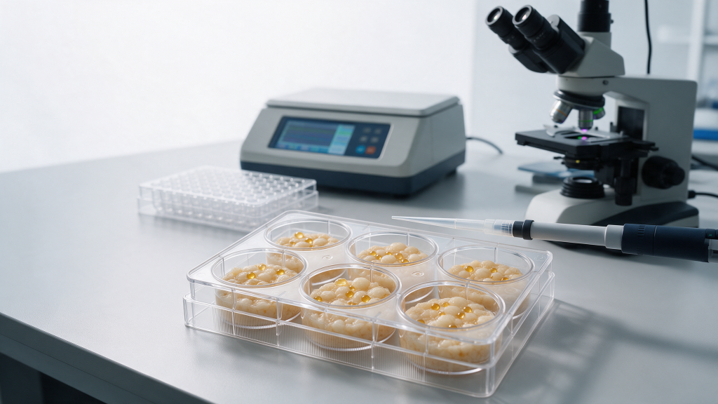
In laboratory terms, fat cell models are systems that copy Adipocyte behavior. These models are used to study lipid metabolism research questions outside a living organism. In-vitro models allow researchers to control nutrient conditions, differentiation signals, and peptide exposure in ways that are difficult in whole organisms. This makes them useful for reproducible, mechanistic experiments.

The most commonly used in-vitro model for adipogenesis is the 3T3-L1 cell line. This line was originally established from mouse embryos and can differentiate into Adipocyte-like cells (Green & Kehinde, 1974; Green & Kehinde, 1975). Because of its reproducible differentiation capacity, 3T3-L1 remains a standard tool in Adipocyte biology.
Differentiation protocols for 3T3-L1 cells have been optimized over time. A commonly cited protocol uses induction media containing insulin, dexamethasone, and isobutylmethylxanthine, followed by insulin-only maintenance media (Zebisch et al., 2012). After differentiation, the cells accumulate lipid droplets and express Adipocyte markers. However, 3T3-L1 cells are not a perfect model of all Adipocyte subtypes. They may display phenotypic characteristics of multiple Adipocyte lineages (Morrison & McGee, 2015). Researchers should therefore choose a model based on the specific pathway or marker being studied.
Primary Adipocyte cultures and other cell lines are also used. Some researchers use human preAdipocyte cell lines to reduce species-related variability. Others use primary stromal vascular fraction cells. Each model has strengths and limitations. The key requirement is that the model is clearly defined and that differentiation is confirmed before experimental treatments begin.
Metabolic peptides are short chains of amino acids studied in metabolic pathways. In Adipocyte research, peptides may be introduced into differentiated fat cell models or into pre-Adipocytes during differentiation. Researchers then measure whether the peptide alters adipogenesis, lipid accumulation, or the expression of metabolic markers.
One peer-reviewed example is the study of glucagon-like peptide-1 (GLP-1) signaling in Adipocyte formation. Research has shown that GLP-1 receptor signaling can regulate Adipocyte formation in vitro (Challa et al., 2012). This demonstrates how a peptide hormone can be examined in an Adipocyte model without implying any effect in humans. The experimental system is the cell culture, and the outcome is a measured change in cellular markers.
In the Elivena catalog, AOD 9604 5mg is presented as a research-use-only product within the metabolic peptides category. AOD9604 has been described in the literature as a synthetic lipolytic domain of human growth hormone used in metabolic studies (Ng et al., 2000). Researchers interested in AOD9604 and Adipocyte models should verify primary studies that directly measure Adipocyte endpoints.
When designing a peptide-Adipocyte experiment, researchers generally include the following steps:
This workflow is a general framework. Specific peptide effects must be established through direct experimental evidence.
Lipid metabolism research depends on measurable markers. In Adipocyte models, researchers commonly evaluate lipid droplet formation, lipolysis, and the expression of genes or proteins involved in lipid handling. These markers allow comparison across experimental conditions.
Lipolysis is a major endpoint. The breakdown of stored triglycerides into free fatty acids and glycerol can be measured in culture media (Lafontan & Langin, 2009). Lipid droplet-associated proteins, such as perilipins, are also used as markers because they coat the surface of lipid droplets and regulate access to stored lipids (Brasaemle, 2007).
Adipocytes are also secretory cells. They release adipokines and other signaling molecules that contribute to metabolic regulation (Frühbeck et al., 2001). Therefore, researchers may measure secreted factors in conditioned media. They may also measure the expression of key transcription factors such as PPARγ and C/EBPα to confirm that the adipogenic program has been activated.
Common variables measured in Adipocyte peptide research include:
This article is intended for laboratory research education only. The compounds discussed are not for human consumption and are not intended for human or animal use. Researchers should verify all original sources and ensure that any experimental product is handled according to applicable institutional and legal requirements.
