Your cart is empty.
- Categories

Cell migration is a basic biological process. In laboratory settings, researchers study how cells move from one location to another. This movement is a critical component of tissue repair models. Regenerative peptides are frequently studied in these in-vitro models. Scientists use cell migration assays to observe and quantify this cellular movement. This article explains how these assays function within peptide research.
Cell migration refers to the physical movement of cells. In a lab, researchers use specific tests to watch this process. These tests are called cell migration assays. They provide measurable data on cellular motility.
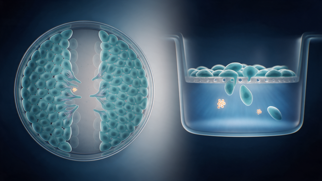
A scratch assay is a very common test. In this test, a scientist grows a layer of cells in a culture dish. The cells grow until they form a complete, single layer. The scientist then uses a tool to make a scratch through the cell layer. This scratch creates an empty, cell-free space. The cells on the edges of the scratch then move to fill this empty area.
This test is also known as a gap-closure assay in vitro . “In vitro” means the test happens in a controlled lab environment. It happens outside a living organism. The gap-closure assay measures how quickly cells close the empty gap. It is a simple and affordable method for studying cell movement.
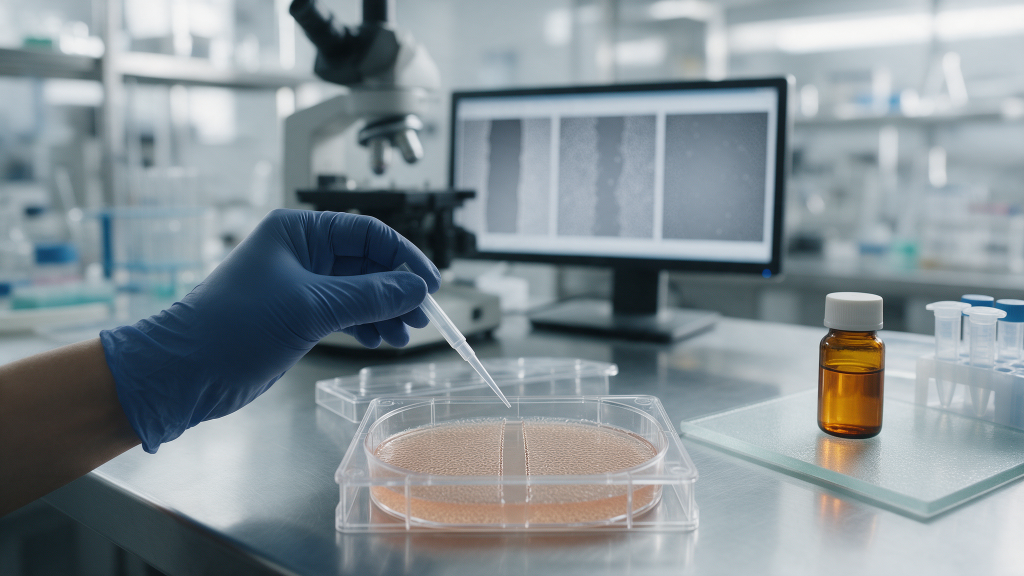
The scratch assay follows a clear set of steps. First, researchers seed the cells in a multi-well plate. They wait for the cells to attach and grow into a full layer. Second, they use a sterile pipette tip to make a straight scratch in each well. Third, they wash the wells to remove any detached cells. This ensures the gap is completely clear. Fourth, they add the culture medium containing the test peptide. Finally, they place the plate under a microscope. They take photos at regular intervals over several hours.
Data analysis involves comparing the gap size at time zero to the gap size at later time points. Researchers use image analysis software. The software traces the edges of the migrating cells. This provides objective and accurate data.
The scratch assay is not the only method. Researchers also use transwell assays. A transwell assay uses a small chamber with a porous membrane. Cells are placed in the upper chamber. A substance that attracts cells is placed in the lower chamber. Migrating cells move through the pores in the membrane. Researchers then count the cells that passed through the membrane. This method separates migration from proliferation naturally. The membrane pores are usually too small for cells to divide and pass through.
In these laboratory tests, researchers track specific data points. What is measured in vitro? Scientists measure the rate of cell movement into the scratch area. They also measure the percentage of the gap that closes over time.
First, researchers measure the migration rate. This is the speed at which cells move into the empty space. A faster migration rate means the cells are moving more quickly. Scientists calculate this by measuring the distance the cells travel over a set period of time.
Second, researchers measure the gap closure percentage. This is the amount of the scratch that the cells have covered at a given time. If the gap is fully closed, the percentage is one hundred. Researchers take images of the cells at regular intervals. They use software to measure the remaining gap area.
Third, researchers must separate cell migration from cell proliferation. Cell proliferation is the process where cells divide and create new cells. Both migration and proliferation can close a gap. Scientists use special controls to ensure they measure only migration. They might use a substance that stops cell division. Mitomycin C is a common substance used for this purpose. This ensures the gap closes only because cells moved, not because they multiplied.
Regenerative peptides are short chains of amino acids. Researchers study them to understand their properties in tissue repair models . More information is available on the regenerative peptides category page. These peptides are studied for their interactions with cellular proteins.
One example is TB-500. TB-500 is a synthetic version of Thymosin Beta-4. It is studied for its role in actin binding. Actin is a protein that forms the cellular skeleton. It helps cells move and change shape. Researchers use the scratch assay to study how TB-500 affects cell movement in a petri dish.
In the lab, scientists add TB-500 to the cell culture. They observe the cells in the gap-closure assay in vitro. They observe whether the peptide changes the migration rate. They do not study human outcomes or therapeutic effects . They only study the cellular mechanics in a controlled environment. Visit the TB-500 product page to learn more about this compound.
Related research areas include TB-500 and Angiogenic Pathway Research. Another related topic is Fibroblast Activity Models in Peptide Research. These models help scientists understand peptide interactions at the cellular level. Angiogenic pathway research studies how new blood vessels form in vitro. Fibroblast models study the cells that create connective tissue.
This information is strictly for laboratory professionals. All compounds discussed here are for in-vitro research only. They are not for human consumption. They are not for veterinary use. Researchers use these peptides to study cellular behavior in controlled settings. No therapeutic benefits are implied. No dosing guidelines for humans are provided. The research focuses entirely on what the compound is and what is studied in the lab .
For research use only. This content is provided for in-vitro laboratory research purposes. The compounds discussed are not for human consumption and are not approved by the FDA or EMA to prevent, treat, or cure any condition. Introduction into humans or animals is prohibited by law. References:
