Your cart is empty.
- Categories

The extracellular matrix (ECM) is a network of proteins and carbohydrates that surrounds cells in tissues. It provides structural support and chemical signals that guide cell behaviour. Regenerative peptides are short chains of amino acids. They are studied in ECM research for their possible roles in tissue repair processes. This article reviews the main research themes, peptide candidates, and laboratory methods used to study regenerative peptides in ECM models [1]. All content is for research use only.
The ECM is not an inert scaffold. It is a dynamic environment. Researchers study the ECM because it influences cell shape, differentiation, and migration. The main components of the ECM include collagen, elastin, fibronectin, laminin, and proteoglycans. Collagen is the most abundant protein in the ECM. It gives tissues tensile strength. Elastin allows tissues to return to their original shape after stretching. Fibronectin and laminin are adhesive glycoproteins. Proteoglycans are proteins with attached carbohydrate chains. They help the matrix hold water and resist compression.
ECM turnover is the continuous process of breaking down and rebuilding ECM components. In simple terms, cells remove old matrix material and produce new matrix material. This balance is important in tissue maintenance and repair. Regenerative peptides are studied to understand whether they influence ECM turnover signals in laboratory models. Researchers do this by measuring gene expression, protein levels, and matrix organisation.
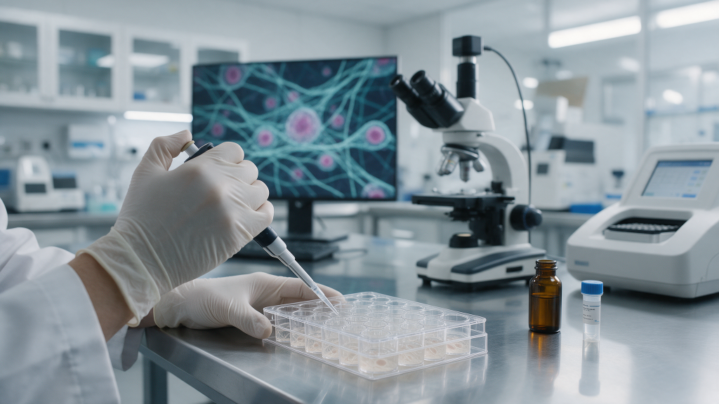
Several regenerative peptides are examined in ECM-related experiments. These peptides are short synthetic sequences of amino acids. They are not nutrients or medications. They are laboratory tools used to test hypotheses about cell-matrix interactions.
BPC-157: BPC-157 is a synthetic peptide composed of 15 amino acids. It is derived from a protein found in gastric juice. In laboratory models, BPC-157 is often used to study fibroblast activity, cell migration, and collagen-associated gene expression. Fibroblasts are connective tissue cells that produce collagen and other ECM proteins. Researchers may introduce BPC-157 into cell cultures to observe whether matrix-related genes change. The BPC-157 10 mg product page provides compound specifications for laboratory reference.
TB-500: TB-500 is a synthetic fragment of thymosin beta-4. Thymosin beta-4 is an actin-binding protein. Actin is a protein that helps cells move and maintain shape. In ECM research, TB-500 is studied in cell migration assays and matrix remodelling models. The TB-500 product page offers reference details for in-vitro studies.
Copper peptides: Copper peptides are small peptide complexes that bind copper ions. They are used in fibroblast models to examine collagen expression. Collagen expression is the production of collagen protein from collagen genes. These models help researchers understand how peptide-copper complexes may affect matrix synthesis signals.
Collagen-associated gene expression refers to the activity of genes that produce collagen and related matrix proteins. In simple terms, researchers measure whether cells read collagen genes more or less after exposure to a compound. This is done in cell culture. Common methods include quantitative polymerase chain reaction (qPCR) and RNA sequencing. To ensure accuracy and reproducibility, qPCR experiments in these models are typically designed following the MIQE guidelines (Bustin et al., 2009). These data help researchers compare gene activity between compound-exposed cells and control cells.
Regenerative peptides are often tested for their influence on collagen type I and type III expression in fibroblasts. Type I collagen is the main collagen in skin, tendon, and bone. Type III collagen is found in early wound healing and in more elastic tissues. Changes in these gene markers are used as laboratory endpoints. They do not prove a clinical effect.
Matrix remodelling is the structural reorganisation of the ECM. In simple terms, it is how cells reshape and replace matrix components. This process involves enzymes such as matrix metalloproteinases (MMPs). MMPs are proteins that break down ECM components. Tissue inhibitors of metalloproteinases (TIMPs) slow down this breakdown. Researchers study the balance between MMPs and TIMPs to understand ECM turnover. Regenerative peptides may be assessed for their ability to shift this balance in cell culture models.
Common measurements include MMP activity assays and protein level analysis. These experiments help establish whether a peptide changes the rate of matrix breakdown or rebuilding. They are purely observational and research-oriented.
Several laboratory methods are used to study regenerative peptides in ECM research. Each method provides a different type of information.
Fibroblast activity models: Fibroblasts are cultured in dishes or on scaffolds. A peptide is added to the culture medium. Researchers then measure cell proliferation, viability, or matrix protein production. These models are foundational to in vitro research and are standardized in core cell culture protocols (Freshney, 2010).
Cell migration assays: A cell migration assay measures how cells move across a surface or through a barrier. In simple terms, researchers create a gap in a cell layer and record how quickly cells close the gap. The in vitro scratch assay is a widely used, inexpensive method for this type of analysis (Liang et al., 2007). This method is relevant to ECM research because cell movement is essential for tissue repair. TB-500 and BPC-157 are often included in these assays.
Gene expression analysis: Researchers extract RNA from cells after peptide exposure. They then measure specific matrix-related genes. This method shows whether gene activity changes at the molecular level, with qPCR practices guided by established minimum information standards (Bustin et al., 2009).
Protein quantification: Enzyme-linked immunosorbent assay (ELISA) and Western blot are common methods to measure protein amounts. ELISA is a plate-based test that detects a specific protein using antibodies (Engvall & Perlmann, 1971). Western blot separates proteins by size and identifies them with antibodies (Mahmood & Yang, 2012). These methods confirm whether changes in gene expression lead to changes in protein levels.
Matrix staining and imaging: Staining methods such as sirius red or Masson’s trichrome can visualise collagen deposition. Picrosirius red staining combined with polarization microscopy is a specific and highly effective method for detecting and differentiating collagen types in the ECM (Junqueira et al., 1979), while Masson’s trichrome provides general connective tissue visualization (Ross & Pawlina, 2020). Researchers then use microscopy to compare matrix content between groups.
Understanding ECM dynamics is a foundation of regenerative medicine research. Regenerative peptides are studied not as therapies but as molecular tools. They may help researchers identify pathways involved in matrix production and turnover. However, results from cell culture cannot be directly translated to whole organisms. Laboratory models control for variables such as cell type, peptide concentration, and exposure time. This control allows researchers to isolate specific molecular effects.

All compounds described in this article are for laboratory and in-vitro research use only. They are not intended for human or veterinary use. They are not drugs, supplements, or therapeutic agents. The information here describes what these peptides are and how they are studied. It does not describe effects in humans. It does not provide dosing, treatment, or outcome information. Researchers should handle all peptides according to institutional safety guidelines. Product pages on Elivena provide compound specifications for laboratory procurement.
This article is for educational purposes in an academic or institutional research context. It does not constitute medical or scientific advice. Statements have not been evaluated by regulatory authorities. Use appropriate personal protective equipment and follow local regulations.
The extracellular matrix is a central research focus in regenerative science. Regenerative peptides such as BPC-157, TB-500, and copper peptides are studied in fibroblast models, cell migration assays, and gene expression experiments. These models help researchers ask precise questions about ECM turnover and collagen-associated gene expression. They do not provide clinical proof. The field continues to develop through controlled laboratory investigation. To explore the peptide category, visit the regenerative peptides collection. To review specific compounds, see the BPC-157 product page and the TB-500 product page.
Closing disclaimer: This article is for research-use-only informational purposes. All products are intended for laboratory and in-vitro research only. They are not for human or veterinary use. Statements about compounds have not been evaluated by regulatory authorities. This content does not provide medical advice or recommend any human use.
