Your cart is empty.
- Categories

Thymosin Beta 4 is an actin-binding peptide studied in laboratory models of cellular movement, tissue organization, and angiogenic signalling. Its research profile makes it relevant to investigators examining pathways involved in regenerative biology.
This article provides a research-use-only overview of Thymosin Beta 4, its relationship to TB-500, and its use in in-vitro peptide study models.
Thymosin Beta 4 is a naturally occurring peptide that binds to Actin. Actin is a structural protein that helps cells maintain their shape and move through their surrounding environment.
The peptide is commonly examined in cell-based research because actin organization is closely linked to cell migration. Cell migration is the movement of cells from one location to another. This process is relevant to laboratory studies of wound-model systems, tissue organization, and cellular repair pathways.
Thymosin Beta 4 is also investigated in models involving angiogenic signalling. Angiogenesis is the formation of new blood vessels from existing vessels. In laboratory research, angiogenic models help investigators evaluate how cells behave under conditions associated with vascular development.

Thymosin Beta 4 and TB-500 are related but should not be treated as identical compounds.
Thymosin Beta 4 refers to the full peptide sequence. TB-500 generally refers to a synthetic fragment related to Thymosin Beta 4. The exact identity, sequence, formulation, and research characteristics can differ between materials described using these names.
This distinction is important when comparing research records, protocols, analytical data, or product documentation. Investigators should review the stated compound identity and available quality information rather than assuming that all materials labelled TB-500 are equivalent to full-length Thymosin Beta 4.
Thymosin Beta 4 is used in several types of laboratory models. These models allow researchers to examine cellular behavior under controlled conditions without making conclusions about human use.
Cell migration assays are laboratory tests that measure how cells move across a surface or through a membrane. These assays can be used to examine changes in cell distribution, movement patterns, or directional migration.
In an in-vitro peptide study, researchers may compare untreated samples with samples exposed to Thymosin Beta 4 under defined experimental conditions. Variables can include cell type, culture medium, exposure duration, concentration range, and assay design.
Results from these models are limited to the conditions tested. They do not establish how the compound behaves in a human or animal system.
Angiogenic models are laboratory systems used to study processes associated with blood-vessel formation. Common approaches may involve cultured endothelial cells, three-dimensional matrix systems, or other controlled assays.
Endothelial cells line the interior surface of blood vessels. Their movement, organization, and response to experimental conditions can be measured in model systems designed to represent selected aspects of angiogenic activity.
Thymosin Beta 4 may be included in these models when researchers are studying the relationship between actin organization, cell movement, and vascular-pattern formation. The purpose is to characterize cellular responses in a defined research setting.
Some regenerative research models use three-dimensional matrices or tissue-like scaffolds. A matrix is a support structure that surrounds cells and influences how they attach, move, and organize.
Three-dimensional systems can provide a more complex environment than flat cell culture. They may be used to examine cellular distribution, structural organization, and interactions between cells and their surrounding matrix.
These models remain experimental. They are designed to isolate specific biological questions and should not be interpreted as evidence of a clinical application.
Actin-binding describes the interaction between a peptide and actin proteins inside cells. Actin exists in different forms and can assemble into filaments. These filaments contribute to the internal framework that supports cell shape and movement.
Researchers study actin-binding peptides because changes in actin organization can affect how cells spread, attach to surfaces, and migrate. These characteristics can be measured using microscopy, staining methods, image analysis, and other laboratory techniques.
A typical study may assess:
The value of these measurements depends on the experimental design, controls, replication, and analytical methods. A single assay does not provide a complete description of a compound’s biological profile.
Thymosin Beta 4 is of interest in regenerative research because it can be examined across several connected areas of cell biology. These include actin regulation, cell migration, matrix interaction, and angiogenic signalling.
Researchers may use the peptide when investigating:
These studies can help define mechanisms and experimental relationships. They do not, by themselves, establish safety, efficacy, approved use, or a therapeutic outcome.
Thymosin Beta 4 can be considered alongside other materials in the regenerative peptides category when planning a broader research-screening strategy.
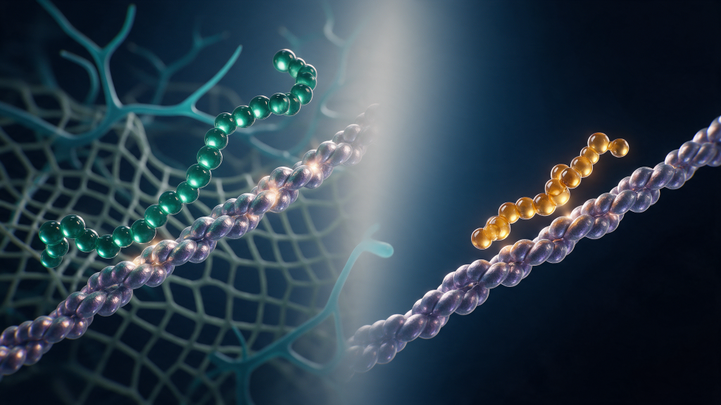
Research teams evaluating Thymosin Beta 4 should begin by confirming the identity of the material under consideration. This includes reviewing the product name, stated sequence, salt or acetate form, quantity, purity information, and available analytical documentation.
The experimental plan should also define:
For product-specific information, researchers can review Thymosin Beta 4 (TB500) 5mg. A separate TB500/Thymosin B4 acetate 10mg variant may also be relevant when the intended study requires a different listed quantity or formulation. Product documentation should be checked carefully before material is included in a laboratory workflow.
Terminology should remain precise throughout the research record. Full-length Thymosin Beta 4, TB-500, and other related fragments should be documented as distinct materials unless analytical evidence supports a specific equivalence.
Thymosin Beta 4 is a subject of laboratory investigation in models involving actin binding, cell migration, and angiogenic signalling. Its use in these models can support controlled research into cellular structure and movement.
Interpretation should remain limited to the model, conditions, and measurements used. In-vitro findings should not be presented as evidence of human or animal outcomes.
For research use only. This content is provided for in-vitro laboratory research purposes. The compounds discussed are not for human consumption and are not approved by the FDA or EMA to prevent, treat, or cure any condition. Introduction into humans or animals is prohibited by law.
1. Safer D, Elzinga M, Nachmias VT. Thymosin beta 4 and Fx, an actin-sequestering peptide, are indistinguishable. J Biol Chem. 1991;266(7):4029-4032.
2. Hertzog M, van Heijenoort C, Didry D, et al. The beta-thymosin/WH2 domain: structural basis for the switch from inhibition to promotion of actin assembly. Cell. 2004;117(5):611-623.
3. Malinda KM, Goldstein AL, Kleinman HK. Thymosin beta 4 stimulates directional migration of human umbilical vein endothelial cells. FASEB J. 1997;11(6):474-481.
4. Koutrafouri V, Leondiadis L, Avgoustakis K, et al. Effect of thymosin peptides on the chick chorioallantoic membrane angiogenesis model. Biochim Biophys Acta. 2001;1568(1):60-66.
5. Sosne G, Christopherson PL, Barrett RP, Fridman R. Thymosin-beta4 modulates corneal matrix metalloproteinase levels and polymorphonuclear cell infiltration after alkali injury. Invest Ophthalmol Vis Sci. 2005;46(7):2388-2395.
6. Goldstein AL, Hannappel E, Sosne G, Kleinman HK. Thymosin beta4: a multi-functional regenerative peptide. Basic properties and clinical applications. Expert Opin Biol Ther. 2012;12(1):37-51.
