
 Best Sellers
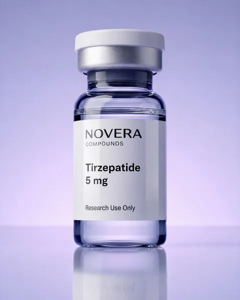 Metabolic
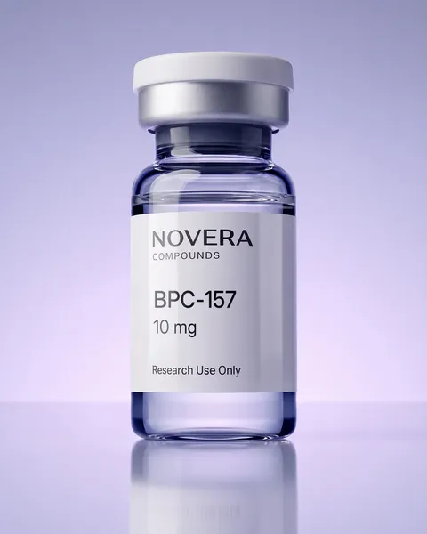 Regenerative
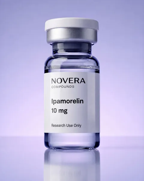 Secretagogues
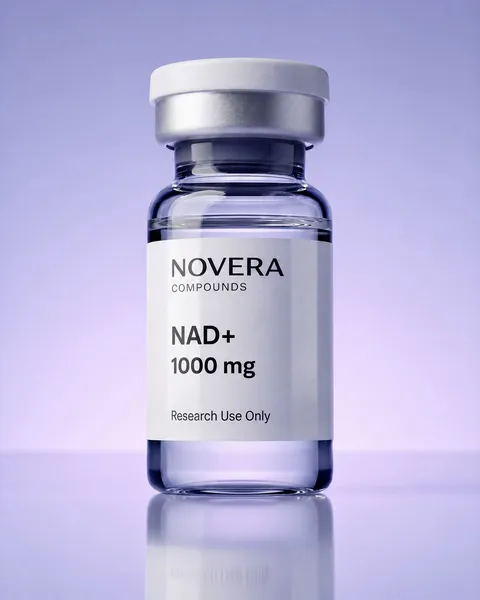 Bioregulators
 Neuropeptides






Thymic peptides are short chains of amino acids studied in relation to the thymus, the organ where T cells mature. T cells are white blood cells that recognize specific molecular targets. In laboratory research, thymic peptides are investigated to understand processes associated with cell development, differentiation, and communication.
This overview is intended for immunology researchers qualified to handle research-grade compounds. It explains how thymic peptides are studied in T-cell differentiation models. The discussion is limited to in-vitro systems, meaning experiments conducted outside a living organism, such as cell cultures and laboratory assays.

What Thymic Peptides Are
A peptide is a chain of amino acids linked together and shorter than a full protein. Thymic peptides are studied in relation to thymus biology and immune-cell development. Researchers examine them as experimental compounds in cell cultures and biochemical assays.
Examples discussed in the research literature include thymosin alpha-1, thymulin, and thymopoietin. These peptides have different structures and research histories, so findings about one should not automatically be applied to another. Researchers investigate them to explore signaling pathways and developmental processes under controlled conditions.
T-Cell Differentiation: The Core Concept
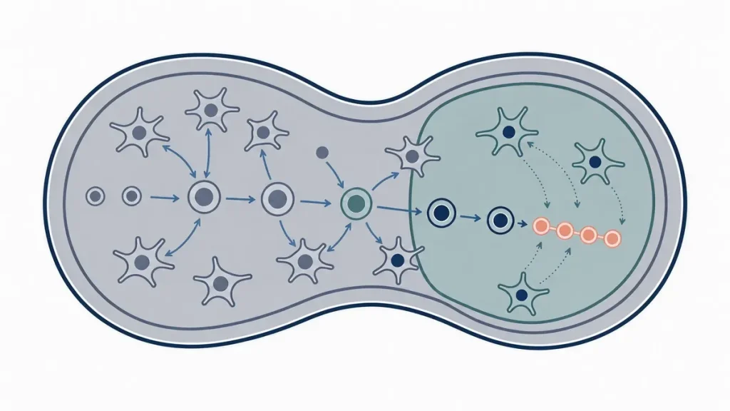
T-cell differentiation is the process by which immature precursor cells develop into distinct T-cell subsets. A precursor is an early-stage cell that has not yet acquired a specialized role. During differentiation, changes in gene activity help cells develop characteristics associated with particular T-cell types.
In the thymus, precursors pass through developmental stages marked by changes in surface markers. A surface marker is a molecule on a cell’s exterior that helps identify its type or stage. Researchers track these markers to map cellular development.
In-vitro models reproduce selected aspects of these stages. They allow researchers to examine associations between individual molecules, including thymic peptides, and cellular development. However, simplified laboratory systems may not reproduce every feature of the thymus.
Thymic Epithelial Cells and the Thymic Microenvironment
Thymic epithelial cells are specialized cells that form part of the thymus’s structure and help shape its internal environment. The two major groups are cortical and medullary thymic epithelial cells. They occupy different regions and provide distinct signals to developing T cells.
Researchers study these cells because they support T-cell differentiation through contact-dependent signals and soluble factors. A soluble factor is a molecule released into surrounding fluid that can interact with nearby cells.
In co-culture experiments, thymic epithelial cells are grown alongside precursor cells to reproduce selected features of the thymic micro-environment. This approach helps researchers investigate cell interactions under defined conditions. Results depend on the cell types, culture conditions, and experimental design.
Immune Signaling in Thymic Development
Immune signaling is the process by which cells send and receive molecular messages. Cytokines are small proteins involved in communication between cells. When a cytokine binds to its corresponding receptor, it can initiate events inside the cell.
A receptor is a protein that recognizes and binds a particular molecule. The interaction may activate an intra-cellular signaling pathway, a sequence of molecular events within a cell. Researchers study these pathways to understand how cellular processes are coordinated.
In thymic peptide research, investigators may measure signaling markers or analyze cytokines in culture medium, the liquid surrounding cells. These measurements help characterize changes associated with experimental conditions. A change in a marker does not, by itself, establish a mechanism or prove that a peptide directly caused the change.
Lymphocyte Models Used to Study Thymic Peptides
A lymphocyte is a type of white blood cell; T cells are one category of lymphocyte. Lymphocyte models in peptide research are in-vitro systems used to investigate cellular processes. Researchers select a model based on the question being studied.
Common approaches include:
- Cell lines. Immortalized cell lines can continue dividing under suitable conditions. They offer consistency but may not reflect all features of cells in native tissue.
- Primary cell cultures. These use cells collected from tissue and maintained in culture. They may retain native characteristics more closely, but can vary between preparations.
- Thymic organ cultures. Small pieces of thymic tissue are maintained in laboratory conditions, preserving some cell contacts and tissue organization.
- Co-culture systems. Precursor cells are grown with thymic epithelial cells or other relevant populations to investigate interactions between cell types.
Each model has limitations. Cell lines are consistent but simplified, while primary cultures may be more representative but harder to standardize. Organ cultures preserve some tissue features without reproducing the full conditions of an intact thymus. Comparing results across models can help researchers assess whether observations hold under different conditions.
Cytokine Expression Assays in Thymic Peptide Research
Cytokine expression assays are laboratory methods used to measure cytokine-related gene activity or protein levels. Gene expression is the process by which information in a gene is used to produce a functional product, such as a protein.
Researchers can measure messenger RNA (mRNA), which carries genetic instructions used in protein production, or measure proteins with methods such as enzyme-linked immunosorbent assays (ELISAs). ELISAs use antibodies, proteins that bind specific target molecules, to detect and quantify substances in a sample.
These methods help characterize the output of a culture system. Researchers may compare control cultures with cultures exposed to a defined experimental condition to identify changes in measured markers. However, differences in cytokine levels do not necessarily show that a peptide directly caused the change. Cell composition, culture conditions, and measurement variability can also affect results. Appropriate controls and complementary assays help researchers interpret the data.
For more detail, see cytokine expression assays in thymic peptide research.
Thymosin Alpha-1 as a Reference Compound
Thymosin Alpha-1 is a thymic peptide investigated in laboratory research involving immune-related signaling and cellular processes. In an experiment, a reference compound may provide a point of comparison between conditions or help evaluate an assay. Its suitability depends on the research question and supporting evidence.
Researchers may examine Thymosin Alpha-1 alongside cytokine expression assays and lymphocyte models to investigate selected cellular markers. Its inclusion does not make it an appropriate reference for every system. Researchers should consider the compound’s characteristics, assay design, and available evidence when interpreting findings.
See the Thymosin Alpha-1 research peptide page for compound-specific information, and the thymic peptides category for related research materials.
Why This Is Studied
T-cell differentiation is an important area of cellular immunology because the thymus supports the development of diverse T-cell populations. Thymic peptides are one area of investigation for researchers examining molecular processes associated with thymus biology.
In-vitro models let researchers compare defined experimental conditions and measure changes in cellular markers, gene expression, or protein levels. This can help isolate associations that are difficult to examine in more complex biological systems. Findings still depend on model quality and experimental design, and results from a cell line or tissue culture may not apply to other models.
Consistent methods and transparent reporting help laboratories compare assays, culture protocols, and cell models. Replication, suitable controls, and independent evidence are important for assessing whether observations are reliable.
Related reading includes Thymosin Alpha-1 in immune research models and lymphocyte models in peptide research.
For Research Use Only
The thymic peptides discussed here are considered solely in the context of in-vitro and laboratory research. This overview does not establish clinical applications, therapeutic effects, or safety for human or animal use. Findings should be interpreted according to the experimental model and available evidence.
Qualified research personnel should follow institutional procedures, laboratory safety requirements, and applicable regulations when handling research-grade compounds. Appropriate training, personal protective equipment, and disposal procedures are essential. For research use only. This content is provided for in-vitro laboratory research purposes. The compounds discussed are not for human consumption and are not approved by the FDA or EMA to prevent, treat, or cure any condition. Introduction into humans or animals is prohibited by law.
References
- Rothenberg EV, Moore JE, Yui MA. Launching the T-cell-lineage developmental programme. Nat Rev Immunol. 2008;8(1):9-21.
- Dardenne M. Zinc and immune function. Eur J Clin Nutr. 2002;56(Suppl 3):S20-S23.
- Savino W, Dardenne M. Neuroendocrine control of thymus physiology. Endocr Rev. 2000;21(4):412-443.
- Rothenberg EV. Transcriptional control of early T and B cell developmental choices. Annu Rev Immunol. 2014;32:283-321.
- Petrie HT, Zúñiga-Pflücker JC. Zoned out: functional mapping of stromal signaling microenvironments in the thymus. Annu Rev Immunol. 2007;25:649-679.
- Love PE, Bhandoola A. Signal integration and crosstalk during thymocyte migration and emigration. Nat Rev Immunol. 2011;11(7):469-477.
- Anderson G, Takahama Y. Thymic epithelial cells: working class heroes for T cell development and repertoire selection. Trends Immunol. 2012;33(6):256-263.
- Abramson J, Anderson G. Thymic epithelial cells. Annu Rev Immunol. 2017;35:85-118.
- Anderson MS, Venanzi ES, Klein L, et al. Projection of an immunological self shadow within the thymus by the Aire protein. Science. 2002;298(5597):1395-1401.
- Takahama Y. Journey through the thymus: stromal guides for T-cell development and selection. Nat Rev Immunol. 2006;6(2):127-135.
- Romani L, Bistoni F, Montagnoli C, et al. Thymosin alpha 1: an endogenous regulator of inflammation, immunity, and tolerance. Ann N Y Acad Sci. 2007;1112:326-338.
- Radtke F, Wilson A, Stark G, et al. Deficient T cell fate specification in mice with an induced inactivation of Notch1. Immunity. 1999;10(5):547-558.
- Brandstadter JD, Maillard I. Notch signaling in T cell homeostasis and differentiation. Open Biol. 2019;9(11):190187.
- von Freeden-Jeffry U, Vieira P, Lucian LA, McNeil T, Burdach SE, Murray R. Lymphopenia in interleukin (IL)-7 gene-deleted mice identifies IL-7 as a nonredundant cytokine. J Exp Med. 1995;181(4):1519-1526.
- Jenkinson EJ, Anderson G. Fetal thymic organ cultures. Curr Opin Immunol. 1994;6(2):293-297.
- Hare KJ, Jenkinson EJ, Anderson G. In vitro models of T cell development. Semin Immunol. 1999;11(1):3-12.
- Zúñiga-Pflücker JC. T-cell development made simple. Nat Rev Immunol. 2004;4(1):67-72.
- Aydin S. A short history, principles, and types of ELISA, and our laboratory experience with peptide/protein analyses using ELISA. Peptides. 2015;72:4-15.
- Leng SX, McElhaney JE, Walston JD, Xie D, Fedarko NS, Kuchel GA. ELISA and multiplex technologies for cytokine measurement in inflammation and aging research. J Gerontol A Biol Sci Med Sci. 2008;63(8):879-884.
- Bustin SA, Benes V, Garson JA, et al. The MIQE guidelines: minimum information for publication of quantitative real-time PCR experiments. Clin Chem. 2009;55(4):611-622.
- Boehm T, Swann JB. Thymus involution and regeneration: two sides of the same coin? Nat Rev Immunol. 2013;13(11):831-838.