
 Best Sellers
 Metabolic
 Regenerative
 Secretagogues
 Bioregulators
 Neuropeptides









Introduction
Cytokine expression assays are laboratory methods used to detect and quantify cytokines that cultured cells release or produce. A cytokine is a small signaling protein that cells use to communicate with one another. Measuring these signals helps researchers describe the state of a cell population in vitro, meaning in a controlled laboratory environment outside a living organism. In thymic peptide research, cytokine measurements can be assessed alongside cell viability, proliferation, and surface markers. These measurements describe laboratory observations rather than demonstrating immune outcomes. This article outlines the main assay formats, experimental design considerations, and practical limitations. All applications discussed are for in-vitro laboratory research only [1].

What Are Cytokines?
Cytokines are immune-signaling proteins involved in communication between cells. Researchers measure them as markers of cellular activity under defined laboratory conditions.
Cytokine expression can be examined at two levels. Protein-level assays measure cytokines in cell culture supernatant or cell lysate. Supernatant is the liquid surrounding cultured cells, while a lysate contains material released when cells are broken open. Transcript-level assays measure messenger RNA (mRNA), which carries instructions used by cells to make proteins.
How Cytokine Assays Work
Several methods measure cytokine expression. The appropriate method depends on the research question, sample type, and number of targets being examined.
ELISA and Protein Detection
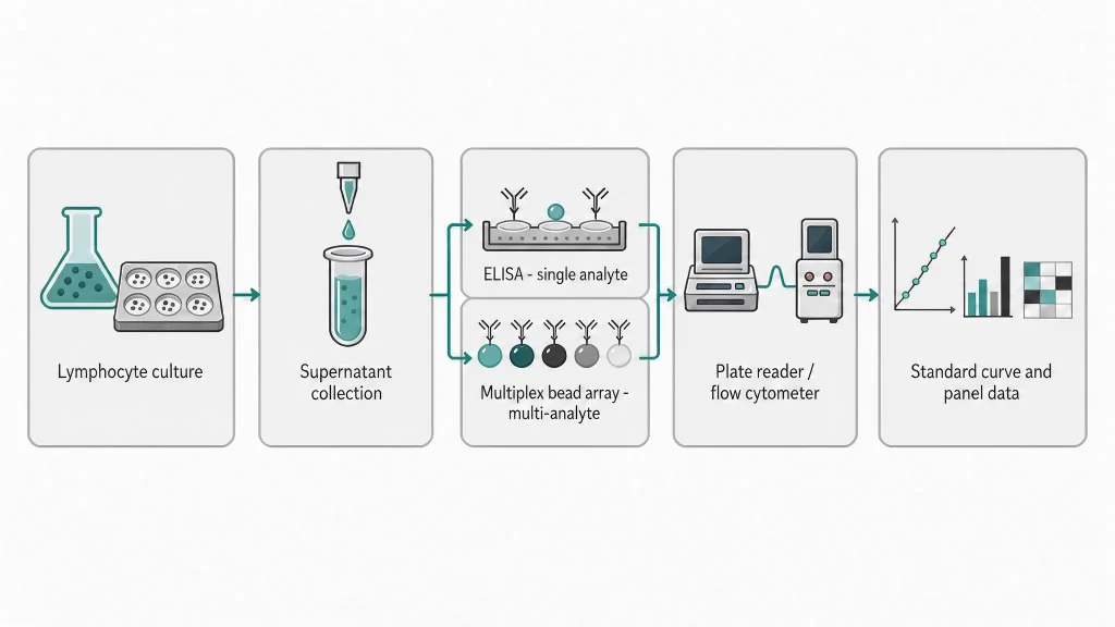
The enzyme-linked immunosorbent assay (ELISA) is a plate-based test that uses antibodies to detect a specific protein. Antibodies are proteins that bind to particular targets.
In a sandwich ELISA, a capture antibody binds the target cytokine, and a second antibody helps detect it. An enzyme then produces a measurable color signal. The signal generally increases with the amount of captured cytokine within the assay’s validated range.
A competitive ELISA uses competition between labeled and unlabeled molecules for antibody-binding sites. Its signal generally decreases as the target concentration increases. This format can be useful when a target is not suited to a two-antibody detection method.
Standard curves help researchers estimate concentrations from measured signals. A standard curve compares known concentrations with their corresponding signals. Dilution checks help identify interference from other sample components. Where possible, researchers should follow the assay manufacturer’s sample-handling instructions and minimize repeated freeze-thaw cycles.
Multiplex Cytokine Profiling and Related Methods
Multiplex cytokine profiling measures several cytokines in a single sample. This approach can conserve sample material, but researchers must validate the panel for cross-reactivity, which occurs when a test detects unintended targets.
Enzyme-linked immunospot (ELISpot) detects cytokine secretion at the individual-cell level. Each visible spot represents a cytokine-secreting cell under the assay conditions.
Intracellular flow cytometry measures cytokines inside cells and identifies the cell populations associated with those measurements. Researchers stain cells for surface markers and intracellular cytokines, allowing them to compare measurements across defined cell subsets.
Transcript-level methods examine gene activity before the corresponding protein accumulates. Reverse transcription quantitative polymerase chain reaction (RT-qPCR) measures selected mRNA targets, while RNA sequencing examines a much broader range of RNA transcripts. These methods require appropriate controls and normalization to support meaningful comparisons.
Application in Thymic Peptide Research
Thymic peptides are short amino acid sequences studied in relation to the thymus, an organ involved in T-cell development. Researchers examine these peptides in controlled laboratory systems, including thymic epithelial cell cultures and lymphocyte models.
Cytokine expression assays can help characterize these systems by measuring selected proteins or transcripts. Commonly measured targets include interleukin-2, interferon gamma, tumor necrosis factor alpha, interleukin-6, and interleukin-10. The appropriate panel depends on the cell model and the research question. These measurements are markers for analysis, not proof of a particular immune effect.
Experimental design influences how the measurements can be interpreted. Unstimulated controls establish baseline measurements, while vehicle controls account for the solvent used in a test preparation. Positive controls help confirm that an assay can detect its intended target. Biological replicates capture variation between independent cultures or donors, while technical replicates help assess measurement consistency.
Sample handling also matters. Researchers should clarify culture supernatants before storage, use aliquots to reduce repeated freeze-thaw cycles, and consider endotoxin contamination when working with sensitive primary cell cultures. Endotoxins are bacterial components that can affect cellular measurements.
For further context, see thymic peptides and T-cell differentiation models and lymphocyte models in peptide research.
Researchers studying these systems may also consult the thymic peptides category and the Thymosin Alpha-1 reference page when identifying compounds relevant to laboratory research.
Reporting and Reproducibility
Consistent reporting helps other researchers understand and reproduce an experiment. Relevant details include assay kit identification, antibody information, standard curve methods, incubation conditions, washing steps, replicate counts, and normalization strategies.
Common technical issues include the hook effect, which can cause falsely low readings when very high target concentrations interfere with antibody-based detection. Matrix effects occur when other sample components alter the measured signal. Cross-reactivity can affect multiplex measurements, while uneven temperature or handling across a plate can introduce variation.
Researchers should follow validated assay protocols, report detection limits, and identify measurements below the limit of quantification. This limit is the lowest concentration that can be measured with acceptable accuracy and precision. For RT-qPCR studies, the MIQE guidelines provide recommendations for transparent reporting.
For Research Use Only
Cytokine expression assays are laboratory tools for measuring proteins and gene transcripts under defined in-vitro conditions. Their measurements should be interpreted within the limits of the assay, sample type, and experimental design. They do not independently establish biological effects or demonstrate clinical outcomes.
For research use only. This content is provided for in-vitro laboratory research purposes. The compounds discussed are not for human consumption and are not approved by the FDA or EMA to prevent, treat, or cure any condition. Introduction into humans or animals is prohibited by law.
REFERENCES
- Sachdeva N, Asthana D. Cytokine quantitation: technologies and applications. Front Biosci. 2007;12:4682-4695. doi:10.2741/2408.
- Tighe PJ, Ryder RR, Todd I, Fairclough LC. ELISA in the multiplex era: potentials and pitfalls. Proteomics Clin Appl. 2015;9(3-4):406-422. doi:10.1002/prca.201400116.
- Dinarello CA. Historical insights into cytokines. Eur J Immunol. 2007;37(Suppl 1). doi:10.1002/eji.200737772.
- Oppenheim JJ. Cytokines: past, present, and future. Int J Hematol. 2001;74(1):3-8. doi:10.1007/BF02982543.
- Miller JFAP. The discovery of thymus function and of thymus-derived lymphocytes. Immunol Rev. 2002;185:7-14. doi:10.1034/j.1600-065X.2002.18502.x.
- Anderson G, Takahama Y. Thymic epithelial cells: working class heroes for T cell development and repertoire selection. Trends Immunol. 2012;33(6):256-263. doi:10.1016/j.it.2012.02.005.
- Klein L, Kyewski B, Allen PM, Hogquist KA. Positive and negative selection of the T cell repertoire: what thymocytes see (and don’t see). Nat Rev Immunol. 2014;14(6):377-391. doi:10.1038/nri3667.
- Goldstein AL, Badamchian M. Thymosins: chemistry and biological properties in health and disease. Expert Opin Biol Ther. 2004;4(4):559-573. doi:10.1517/14712598.4.4.559.
- Romani L, Bistoni F, Gaziano R, et al. Thymosin alpha 1 activates dendritic cells for antifungal Th1 resistance through toll-like receptor signaling. Blood. 2004;103(11):4232-4239. doi:10.1182/blood-2003-10-3509.
- Engvall E, Perlmann P. Enzyme-linked immunosorbent assay (ELISA). Quantitative assay of immunoglobulin G. Immunochemistry. 1971;8(9):871-874. doi:10.1016/0019-2791(71)90454-X.
- Lequin RM. Enzyme immunoassay (EIA)/enzyme-linked immunosorbent assay (ELISA). Clin Chem. 2005;51(12):2415-2418. doi:10.1373/clinchem.2005.051532.
- Vignali DA. Multiplexed particle-based flow cytometric assays. J Immunol Methods. 2000;243(1-2):243-255. doi:10.1016/S0022-1759(00)00238-6.
- Carson RT, Vignali DA. Simultaneous quantitation of 15 cytokines using a multiplexed flow cytometric assay. J Immunol Methods. 1999;227(1-2):41-52. doi:10.1016/S0022-1759(99)00076-7.
- Leng SX, McElhaney JE, Walston JD, Xie D, Fedarko NS, Kuchel GA. ELISA and multiplex technologies for cytokine measurement in inflammation and aging research. J Gerontol A Biol Sci Med Sci. 2008;63(8):879-884. doi:10.1093/gerona/63.8.879.
- Jung T, Schauer U, Heusser C, Neumann C, Rieger C. Detection of intracellular cytokines by flow cytometry. J Immunol Methods. 1993;159(1-2):197-207. doi:10.1016/0022-1759(93)90161-W.
- Lamoreaux L, Roederer M, Koup RA. Intracellular cytokine optimization and standard operating procedure. Nat Protoc. 2006;1(3):1507-1516. doi:10.1038/nprot.2006.231.
- Pala P, Hussell T, Openshaw PJ. Flow cytometric measurement of intracellular cytokines. J Immunol Methods. 2000;243(1-2):107-124. doi:10.1016/S0022-1759(00)00235-0.
- de Jager W, Bourcier K, Rijkers GT, Prakken BJ, Seyfert-Margolis V. Prerequisites for cytokine measurements in clinical trials with multiplex immunoassays. BMC Immunol. 2009;10:52. doi:10.1186/1471-2172-10-52.
- Thavasu PW, Longhurst S, Joel SP, Slevin ML, Balkwill FR. Measuring cytokine levels in blood. Importance of anticoagulants, processing, and storage conditions. J Immunol Methods. 1992;153(1-2):115-124. doi:10.1016/0022-1759(92)90313-I.
- Riss TL, Moravec RA, Niles AL, et al. Cell viability assays. In: Markossian S, Grossman A, Brimacombe K, et al, eds. Assay Guidance Manual. Eli Lilly & Company and the National Center for Advancing Translational Sciences; 2004-. Updated July 1, 2016.
- Tuthill C, Rios I, McBeath R. Thymosin alpha 1: past clinical experience and future promise. Ann N Y Acad Sci. 2010;1194:130-135. doi:10.1111/j.1749-6632.2010.05492.x.